MSS from North Italy, circa 1300, Rome

The text above the picture reads as follows:
MSS from North Italy, circa 1300, Rome
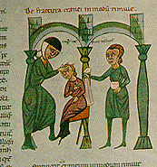
Rolandus Parmensis, Chirurgia, I, 5-6, Surgical examination of a fractured cranium |
“Concerning cranial fracture in the shape of a fissure. Sometimes it chances that the cranium is fractured like a fissure and is split so that neither side seems higher than the other; also that it is uncertain whether the fracture extends inside the cranium. To find out, have the patient hold his mouth and nostrils shut, then blow vigorously; if any breath comes out of the cleft, you know that the cranium is fractured even into the cerebrum. Treat as follows. If the wound is narrow, enlarge it, and unless prevented by bleeding immediately trephine with an iron instrument, very cautiously on both sides of the fissure. Make as many holes as seems wise, then cut the cranium from one whole to another with a bistoury (spathumina), so that the incision extends to the edge of the fissure. Carefully remove pus oozing from above the cerebrum with a silk from a fine linen cloth introduced sideways between the cerebrum and cranium by means of a feather...[then medicate]”. |
| "If there is a stone in the bladder, make sure of it as follows: have a strong person sit on a bench, his feet on a stool; the patient sits on his lap, legs bound to his neck with a bandage, or steadied on the shoulders of the assistants. The physician stands before the patient and inserts two fingers of his right hand into the anus, pressing with his left fist above the patient's pubes. With his fingers engaging the bladder from above, let him work over all of it. If he finds a hard, firm pellet it is a stone in the bladder, which is soft and fleshy; thus you find that it impedes urination; if you want to extract the stone, precede it with a light diet and fasting for two days beforehand, On the third day, having done everything beforehand, as we said, to find whether there is a stone in the bladder, locate the stone, bring it to the neck of the bladder; there, at the entrance, with two fingers above the anus, incise lengthwise with an instrument and extract the stone... [medication, morning and night for nine days]. (Rogerius, Chirurgica, III, 36). |
Manuscript from North Italy circa 1300, Rome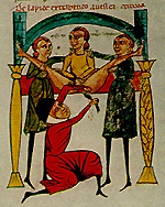
Celsan operation for bladder stones, Rolandus Parmensis, Chirurgia III, 34. |
Manuscript from Byzantium, c. 1100, Florence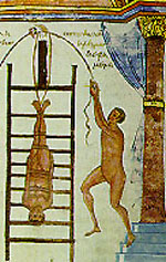
Laurentian Library, MS 74.7, folio 200 Apollonius, Dislocations, 2, in Greek Reduction by Jolting on a ladder |
“Cases where the curvature is low on the spine are best treated with the head downward...Pad the ladder...Lay the patient on it, on his back, using soft but strong bandages, tie his ankles to the ladder; bind his legs together below and above the knees and bandage to the hips. Bandage him loosely at the flanks and chest; tie the arms and hands, extended along his sides, to the body, but not to the ladder. Then raise the ladder against a high tower or house. The ground should be solid ad the assistants well trained so that they will let the ladder fall smoothly and in a vertical position...It is best to drop it from a mast by a pulley...Jolting is best done with such an apparatus, but it is disagreeable to discuss in detail. Cases where the curvature is high up on the spine are better treated with the feet downward...Bind the patient firmly to the ladder at his chest, but loosely at his neck, merely enough to keep it straight. Bring his head to the ladder at the forehead. Bind the rest of the body loosely here and there, only to keep it vertical...Fasten the legs together, but not to the ladder, so they hang in line with the back. |
